
The advancement of science often unfolds through revolutionary paradigm shifts—those enlightening moments when long-held beliefs crumble, making way for fresh perspectives. The latest breakthrough from the Kanso Bioinspired Motion Lab at USC Viterbi School of Engineering epitomizes this process. In their groundbreaking paper published in Nature Physics, titled “Flow physics guides morphology of ciliated organs,” they illuminate the intricate relationship between fluid dynamics and the evolutionary architecture of ciliated tissues. This work is paramount as it transcends traditional classifications and establishes a compelling continuum that ties together the diverse forms of ciliary structures.
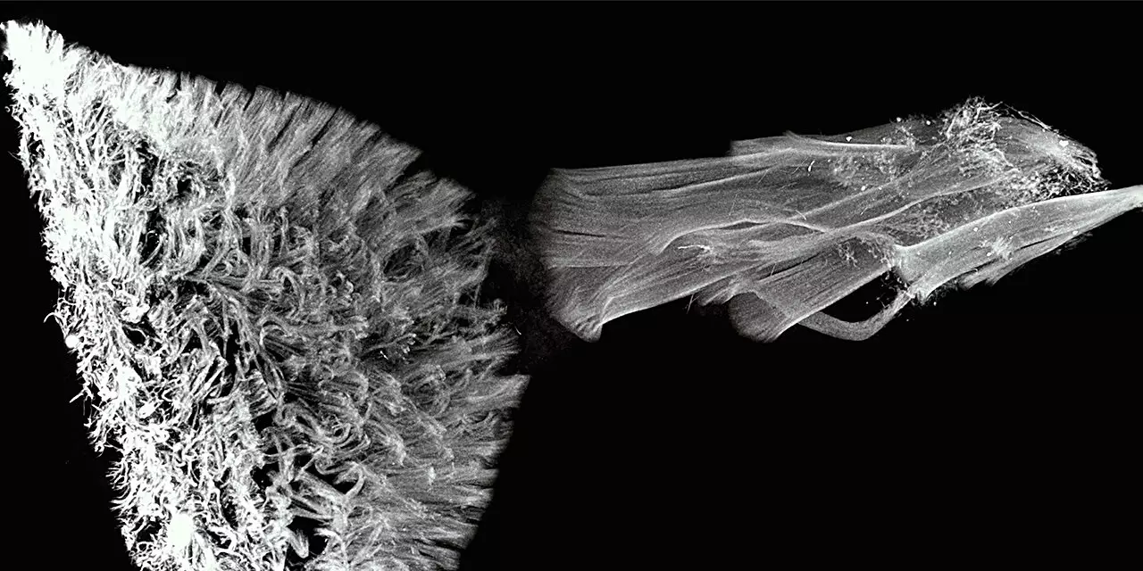
Cilia are vital appendages found within various organisms, playing an essential role in efficiently transporting fluids across different biological systems. Traditionally, the scientific community has classified these structures primarily under two umbrella models: the “carpet” model, characterized by a dense assembly of short cilia, and the “flame” model, defined by longer, parallel cilia that exhibit different movement patterns. This binary approach has persisted largely due to the evolutionary belief that these forms developed independently in response to specific environmental needs. However, the revelations from the Kanso Lab posit that the distinction is less about lineage and much more about mechanical requirements, challenging researchers to re-evaluate existing theories.
A Spectrum of Adaptation
At the core of the Kanso Lab’s findings is a perceptive understanding of how varying physical constraints dictate the functional requirements of ciliated organs. The conundrum is elegantly solved when we recognize that ciliary designs are not merely products of their genetic blueprints but are also shaped by optimized fluid pumping mechanisms. The research conducted by Professor Eva Kanso and her dedicated team, including the talents of doctoral student Feng Ling and research scientist Janna Nawroth, reveals a spectrum that stretches between the two classic models.
This research contributes a universal design framework that allows for these ciliated structures to be categorized based on two primary parameters: lumen diameter and the ratio of cilia to lumen. Such a refined taxonomy not only streamlines our understanding but brings forth a new lexicon for discussing biological design. As the study elucidates, moving along this gradient provides insights into both maximized flow rates and pressure generation—critical for maintaining homeostasis across physiological systems.
Implications for Medical Science
The ramifications of this study resonant far beyond purely academic interest—they hold profound implications for the medical community. The clarity at which the Kanso Lab dissects the relationship between ciliated duct morphology and their pumping capabilities paves the way for novel approaches to treating a myriad of pathologies often linked to cilia dysfunction. Conditions such as bronchiectasis, hydrocephalus, and ectopic pregnancy, which are complex and multifaceted, can now be examined through the lens of these newly discovered design rules.
Furthermore, as modern medicine seeks to understand and combat renal diseases, the principles derived from the morphology of fluid-excreting ciliary flames can serve as exemplary models. Such applications of theoretical research directly translate to clinical insights, enriching our understanding of organ function and informing therapeutic interventions.
Innovative Methodologies in Scientific Inquiry
What stands out in this pioneering research is not just its outcomes, but its innovative approach to scientific inquiry. Conducting experiments in an area fraught with challenges—namely, measuring ciliary beat and fluid flow in situ—is no small feat. Yet, the Kanso Lab expertly combines experimental methods with advanced mathematical modeling to tackle these problems head-on. By redefining the dichotomy between the ‘carpet’ and ‘flame’ models as part of a dynamic continuum, the research simplifies a complex field, offering an intuitive understanding that could have significant ramifications in both engineering and biological studies.
In essence, this work may not only untangle the complexities of ciliary structure and function but also act as a call to arms for future researchers. It serves as a reminder that in science, as in life, discreet categories may shroud a deeper, interconnected reality waiting to be uncovered, and sometimes the most profound insights come from those willing to challenge established norms in pursuit of understanding.